SUMMARY OF HEART STRUCTURE
HEART:-
Heart is a cone shape hollow organ is about 3.0gm in male and 2-5gm in female.
It is 10 cm long. The main function of heart is pumping the blood through the body with the
help of blood vessel.
Position and location of heart: -
The heart is located in the throsis cavity (cheast cavity) in mediastinum (space between lungs).
It is situated more left than right. The base (upper part) of heart is situated behind the strunm, there great vessel that is superior venacava, inferior venacava & coronary sinus enter & leaf.
The apex part of heart is just above the dipharm. This part is situated at the level of fifth interpostal space.
Structure of heart:-
Posterior structure: -
Heart is consist of 3 layers: -
1. Inner layer of heart The Endocardium
2. Middle layer of heart The Myocardium
3. Outer layer of heart The Pericardium
1. Endocardium
It is the inner layer of heart, this layer line the chambers & heart valve. This membrane is smooth & thin that permit the easy movement of blood inside the heart.
2. Myocardium
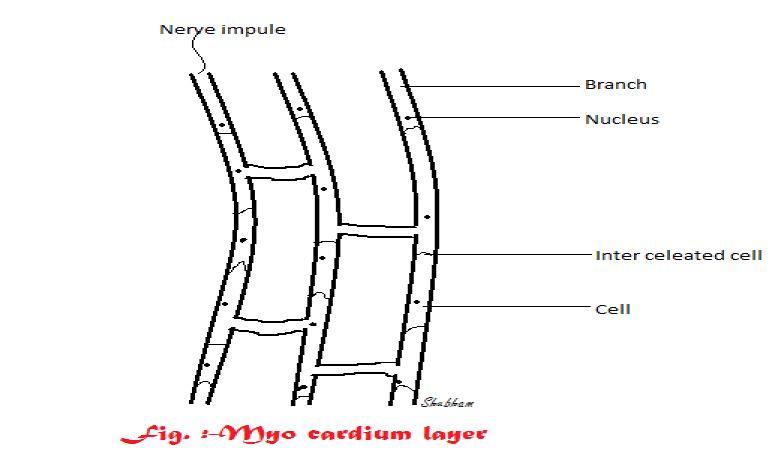
This membrane is also called as heart muscle or cardiac Cell. This part is not under voluntary control.
But when microscopic examination is done they shows cross strips just like skeletal muscle. Each cell has a nucleus or more than one branch. Branches are very close to each other.
Two cells are connected with inter ciliated disc.
These can be seen dark and thick due to this reason myocardium can be seen as a black sheet. Myocardium has cell to cell continuity, so each cell need not to have separate nerve supply, when any impule is insisted.
It separate from cell to cell via branched and intercalated disc over the whole
sheet of muscle (contraction).
Due to this myocardium the artria and ventrical to contract.
The myocardium is thickest of the apex part and thin toward the base.
3. Pericardium

The outer most layer.
This is the outer most layer of heart. Pericardium is consist of two sac-
(i) Outer sac- It is made up by fibrous tissue. It is the toughest part of heart due to this toughness it is prevent the over distension of heart.
(ii) Inner sac- It is term as serus membrane, it consist of flat epithial cells. It secretes serus fluid (pericardial fluid) in between parietal & visceral pericrluin & allow the smooth movement of beat. These two layer are very close to each other.
The perital pericardium is connected with fibrous sac at one end & the other end is connected with the pericardial cavity while the vesal pericardial is connected with pericardial cavity at one end & the other connected with myocardium.
2. Interior structure: -

Heart having four chambers that is two artria & two ventricles. They are consisting of (made up) myocardium & covered by endocardium. Before birth there is a hole in intra artial septum which causes pure blood of mother to fetus enters in right atrium. This blood enter in left atrium threw this hole & this pure blood get enter in left ventricle and supplied to the fetus body threw artria. Because there are no lungs development in fetus but after birth when, the lungs developed this hole is closed and blood cannot cross the septum from one side to another.
Valve of heart : -
Each site that is left or right site of heart having artrioventricular valve or tricuspid
valve having three flaps they divide right artria & ventrical/ left artria & ventrical valve.
They are as mitreal valve/bicuspid valve. They divided left artria & ventricles.
Arotic valve: -
Also called as arotic semilular valve.
These present on junction of left ventrical & arota.
Pulmonary semilular valve: -
These valve present in pulmonary artre that carry impure blood to lungs. It also located
in pulmonary vein that carries pure blood form lungs to left artreum. All the valve have
same function that they prevent the back flow of blood. Tricuspid valve are attach with
capillary muscle by chordatendenae (having 80% collegen and 20% elatives). When
these capillary muscle get relax valve open and on cotarating of these capillary muscle
the valve get close.
Mitrul also having the same type muscle & cordaetendanae having similar function to
that of tricuspid valve.
S.A. Node: -
Sinoatrial node
SA node is situated near the opening of superior venacava at thr right atrium. It is
called as natural pace maker of heart because it has the ability to generate its own
impules.
Myocardium of SA node shows high affinity for sodium ion increases.
Sodium ion concentration intracellulary cause concentration of ………………. And
generation of nerve impules.
A.V.Node: -
It is located near the intra artial septum it recive nerve impulses from S.A. node &
propagate them both the ventricles threw percinfibers.

The main function of heart is to maintain a constant blood circulation throughout the body.
The heart act as a pump and cardiac cycle is also known as heart beat.
That is cardiac cycle is consist of a series of event or we can say that cardiac cycle is simultaneous contraction of two arteria followed by a fraction of second letter by stimulate contraction of two ventricles.
The time duration for complete cardiac cycle is 0.8 sec at the first level both the ventrical get contracted that will causes closing of both the aretio, ventricle valve (tri or bi cuspid valve).
Pushes the blood in lungs & all whole body threw arota this will causes a sound called as LUB (first sound) - This sound is loud and longest, this sound occurs due closing of A.V. valve at the letter stage both the artea get contracted Opening of A.V. valve cecrate another sound called as- DUP sound (second sound) - After this cardiac diastole (filling of heart chambers) for this time is needed 0.4 sec, so,
cardiac cycle is equal to 0.8sec.
Complete cardiac cycle (0.8sec)
= ventrical systole (0.3sec) + artial systole(0.18sec) + complete cardiac cycle(0.4sec)


I Like to add one more important thing here, The Blood Preparation Market is expected to be around US$ 66.80 Billion by 2025 at a CAGR of 4.5% in the given forecast period.
ReplyDeleteThis comment has been removed by a blog administrator.
ReplyDelete